
Expo
view channel
view channel
view channel
view channel
view channel
view channel
view channel
RadiographyMRI
Nuclear MedicineGeneral/Advanced ImagingImaging ITIndustry News
Events

- Cutting-Edge Technology Enhances Chest X-Ray Classification for Superior Patient Outcomes
- AI Model Accurately Estimates Lung Function Using Chest X-Rays
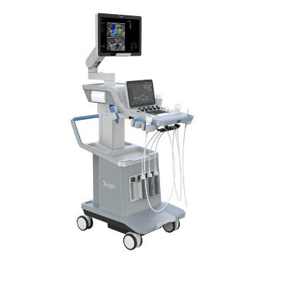
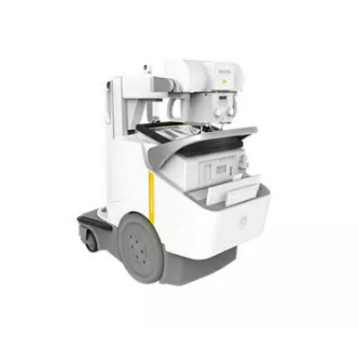
- High-Powered Motorized Mobile C-Arm Delivers State-Of-The-Art Images for Challenging Procedures
- Injury Prediction Rule Reduces Radiographic Imaging Exposure in Children
- AI Detects More Breast Cancers with Fewer False Positives
- AI Outperforms Clinical Tests at Predicting Alzheimer’s Progress from MRI Scans
- Artificial Intelligence Speeds Up Examination of Heart Images from Magnetic Resonance Imaging
- AI Model Diagnoses Traumatic Brain Injury from MRI Scans With 99% Accuracy
- New MR Image Guidance Software Provides Direct Tumor Visualization during HDR Prostate Procedures
- Brain Connectivity on MRI Predicts Parkinson’s Disease Progression
- PET/CT Imaging Using New Tracing Agent Could Become ‘Gold Standard’ Test for Prostate Cancer Detection
- New Imaging Technique Detects Aggressive Lung and Prostate Cancers
- First Specific PET Scan for TB to Improve Treatment
- PET/CT Superior at Lesion Detection for Head and Neck Paragangliomas than Gold Standard MRI
- New Radiotracer Generates High Quality and Readily Interpretable Images of Cardiac Amyloidosis
- Ultrasound Beam Triggers ‘Nanodroplets' For Targeted Drug Delivery
- Ultrasound Technology Breaks Blood-Brain Barrier for Glioblastoma Treatment
- Implantable Ultrasound Device Could Replace Electrodes for Deep Brain Stimulation
- Robotic Ultrasound Systems to Assist Doctors during Surgery
- Functional Ultrasound Imaging Records Brain Activity through Transparent Skull Implant
- Breakthrough Brain PET System Aids Diagnosis of Neurological Disorders
- Ultra-High-Performance PET System Provides Never Before Seen Brain Images
- Artificial Intelligence Tool Enhances Usability of Medical Images
- New AI Tool Accurately Detects Six Different Cancer Types on Whole-Body PET/CT Scans
- Innovative Imaging Technique Helps Assess Bone Loss after Bariatric Surgery
- Global AI in Medical Diagnostics Market to Be Driven by Demand for Image Recognition in Radiology
- AI-Based Mammography Triage Software Helps Dramatically Improve Interpretation Process
- Artificial Intelligence (AI) Program Accurately Predicts Lung Cancer Risk from CT Images
- Image Management Platform Streamlines Treatment Plans
- AI Technology for Detecting Breast Cancer Receives CE Mark Approval
- GE HealthCare Acquires Intelligent Ultrasound Group’s Clinical Artificial Intelligence Business
- Bayer and Rad AI Collaborate on Expanding Use of Cutting Edge AI Radiology Operational Solutions
- Polish Med-Tech Company BrainScan to Expand Extensively into Foreign Markets
- Hologic Acquires UK-Based Breast Surgical Guidance Company Endomagnetics Ltd.
- Bayer and Google Partner on New AI Product for Radiologists
Expo
 view channel
view channel
view channel
view channel
view channel
view channel
RadiographyMRI
Nuclear MedicineGeneral/Advanced ImagingImaging ITIndustry News
Events
Advertise with Us
view channel
view channel
view channel
view channel
view channel
view channel
view channel
RadiographyMRI
Nuclear MedicineGeneral/Advanced ImagingImaging ITIndustry News
Events
Advertise with Us
- Cutting-Edge Technology Enhances Chest X-Ray Classification for Superior Patient Outcomes
- AI Model Accurately Estimates Lung Function Using Chest X-Rays
- High-Powered Motorized Mobile C-Arm Delivers State-Of-The-Art Images for Challenging Procedures
- Injury Prediction Rule Reduces Radiographic Imaging Exposure in Children
- AI Detects More Breast Cancers with Fewer False Positives
- AI Outperforms Clinical Tests at Predicting Alzheimer’s Progress from MRI Scans
- Artificial Intelligence Speeds Up Examination of Heart Images from Magnetic Resonance Imaging
- AI Model Diagnoses Traumatic Brain Injury from MRI Scans With 99% Accuracy
- New MR Image Guidance Software Provides Direct Tumor Visualization during HDR Prostate Procedures
- Brain Connectivity on MRI Predicts Parkinson’s Disease Progression
- PET/CT Imaging Using New Tracing Agent Could Become ‘Gold Standard’ Test for Prostate Cancer Detection
- New Imaging Technique Detects Aggressive Lung and Prostate Cancers
- First Specific PET Scan for TB to Improve Treatment
- PET/CT Superior at Lesion Detection for Head and Neck Paragangliomas than Gold Standard MRI
- New Radiotracer Generates High Quality and Readily Interpretable Images of Cardiac Amyloidosis
- Ultrasound Beam Triggers ‘Nanodroplets' For Targeted Drug Delivery
- Ultrasound Technology Breaks Blood-Brain Barrier for Glioblastoma Treatment
- Implantable Ultrasound Device Could Replace Electrodes for Deep Brain Stimulation
- Robotic Ultrasound Systems to Assist Doctors during Surgery
- Functional Ultrasound Imaging Records Brain Activity through Transparent Skull Implant
- Breakthrough Brain PET System Aids Diagnosis of Neurological Disorders
- Ultra-High-Performance PET System Provides Never Before Seen Brain Images
- Artificial Intelligence Tool Enhances Usability of Medical Images
- New AI Tool Accurately Detects Six Different Cancer Types on Whole-Body PET/CT Scans
- Innovative Imaging Technique Helps Assess Bone Loss after Bariatric Surgery
- Global AI in Medical Diagnostics Market to Be Driven by Demand for Image Recognition in Radiology
- AI-Based Mammography Triage Software Helps Dramatically Improve Interpretation Process
- Artificial Intelligence (AI) Program Accurately Predicts Lung Cancer Risk from CT Images
- Image Management Platform Streamlines Treatment Plans
- AI Technology for Detecting Breast Cancer Receives CE Mark Approval
- GE HealthCare Acquires Intelligent Ultrasound Group’s Clinical Artificial Intelligence Business
- Bayer and Rad AI Collaborate on Expanding Use of Cutting Edge AI Radiology Operational Solutions
- Polish Med-Tech Company BrainScan to Expand Extensively into Foreign Markets
- Hologic Acquires UK-Based Breast Surgical Guidance Company Endomagnetics Ltd.
- Bayer and Google Partner on New AI Product for Radiologists


.")


")
")
")
")
")
")
")
")
")
 are powered by wires (gold) that deliver electrical stimulation (Photo courtesy of MIT)")

")
")
.jpeg "Image: Researchers have developed a more accurate way to scan for TB using PET (Photo courtesy of Adobe Stock)")
")
 scanner (Photo courtesy of Positrigo)")
")
")


")
")
")
")


