




Expo
view channel
view channel
view channel
view channel
view channel
view channel
view channel
RadiographyMRIUltrasoundNuclear Medicine
Imaging ITIndustry News
Events

- AI Tool Predicts Five-Year Breast Cancer Risk from Mammograms
- AI Mammography Tools Detect Early Breast Cancer Signs Years Before Diagnosis
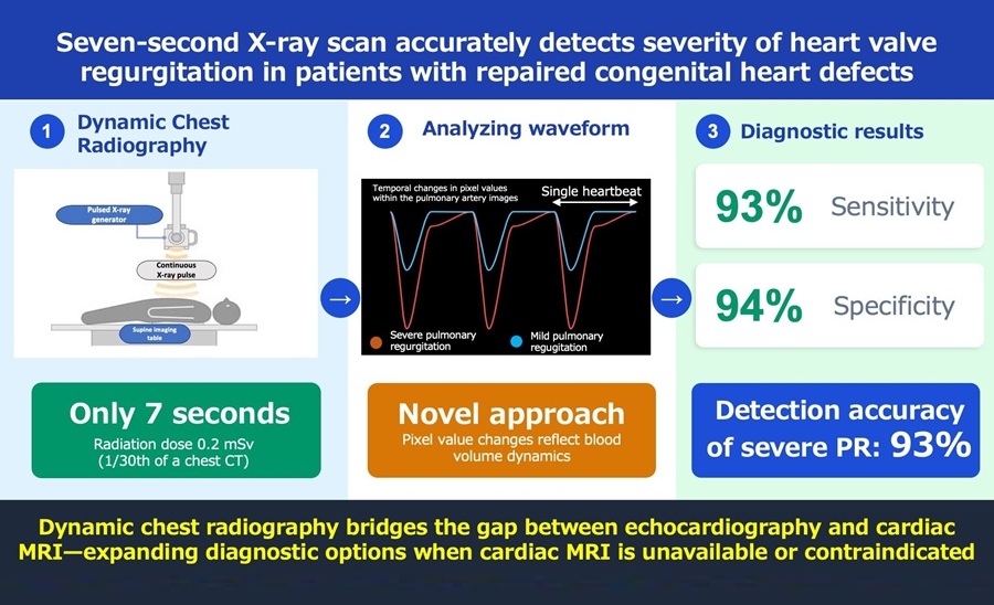
- Rapid X-Ray Test Quantifies Pulmonary Regurgitation After Tetralogy of Fallot Repair
- AI Tool Flags Osteoporosis Risk from Routine Chest X-Rays
- Simple Chest X-Ray Measure Predicts Survival After Lung Cancer Surgery
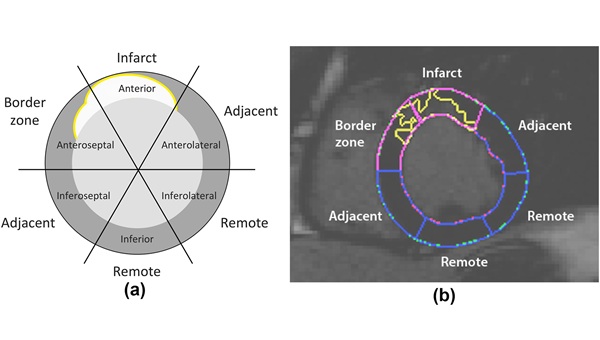
- Cardiac MRI Detects Hidden Heart Dysfunction After Heart Attack
- AI Tool Reveals Cortical Lesions on Standard MRI in Multiple Sclerosis
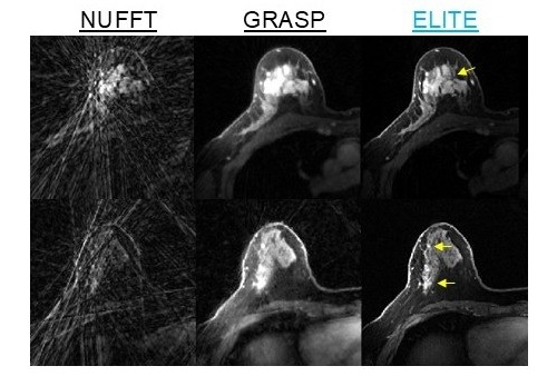
- AI Reconstruction Tool Speeds Dynamic Breast MRI and Improves Cancer Detection
- International Study Assesses AI for Prostate Cancer MRI Interpretation
- AI Approach Could Shorten Advanced Brain MRI Scans by Up to 90%
- Second PSMA PET Identifies Disease in More Than Half of Negative Cases
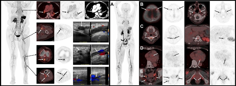
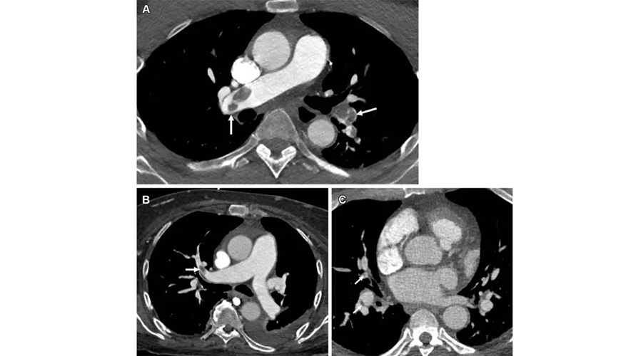
- New PET Tracer Detects DVT and Pulmonary Embolism in One Scan
- Targeted PET Platform Guides Osteosarcoma Resection and Margin Verification
- Portable PET System Enables Real-Time Bedside Guidance for Biopsies and Ablations
- AI Model Predicts Radiation Dose Before Prostate Cancer Therapy
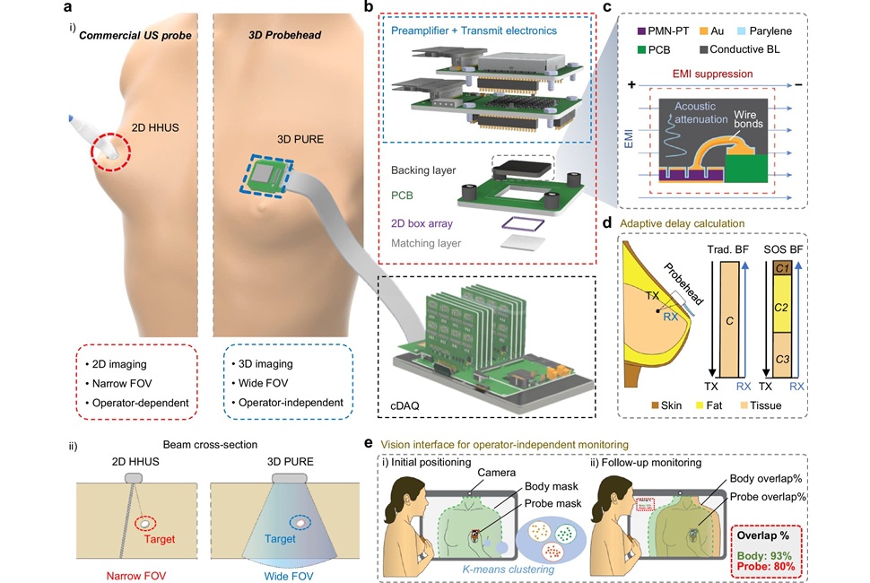
- Portable 3D Ultrasound System Enables Reproducible Breast Cancer Monitoring
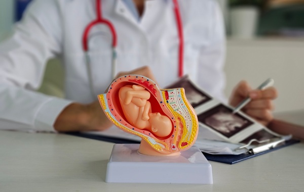
- AI Ultrasound Platform Receives CE Mark for Prenatal Screening
- Hybrid Imaging Platform Reveals How Sleep Supports Brain Waste Removal
- AI Robotic Ultrasound System Automates Echocardiography and Improves Consistency
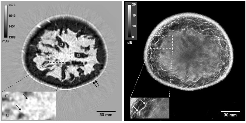
- Whole Cross-Section Ultrasound System Enables Operator-Independent Imaging
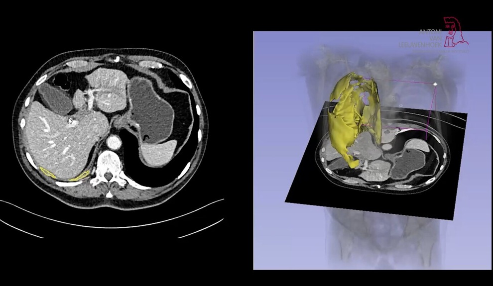
- Virtual Staining Technique Creates Histology Images from CT Data
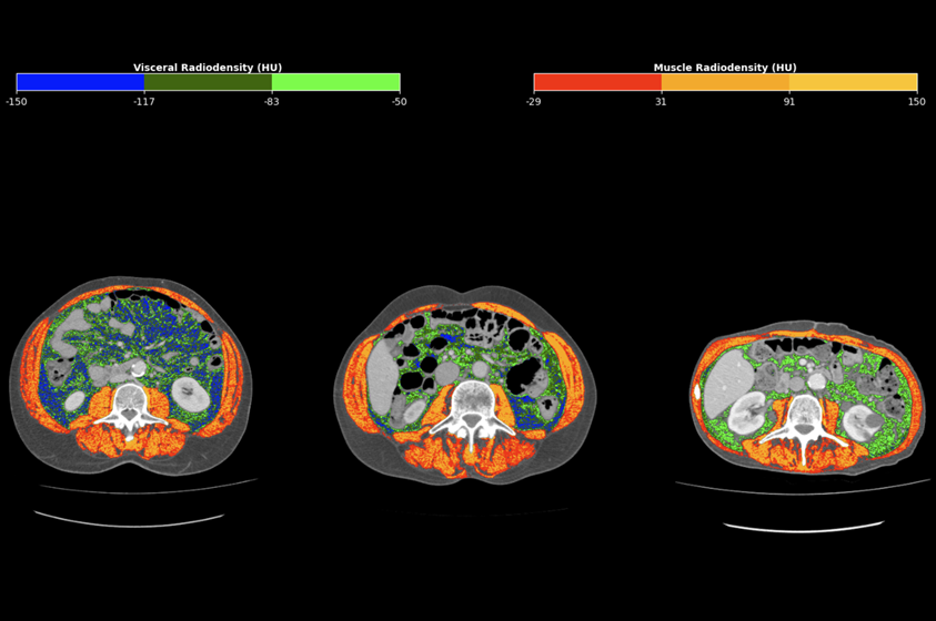
- CT-Derived Biomarker Predicts Outcomes in Gastric Cancer
- AI Tool Enhances Response Assessment and Survival Prediction in Pleural Mesothelioma
- AI Tool Enables Real-Time Diffuse Optical Tomography for Brain Lesion Detection
- New SPECT/CT Method Differentiates Inflammation from Fibrosis in Interstitial Lung Disease
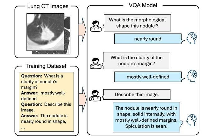
- Interactive AI Tool Supports Explainable Lung Nodule Assessment
- Breast Imaging Software Enhances Visualization and Tissue Characterization in Challenging Cases
- New Google Cloud Medical Imaging Suite Makes Imaging Healthcare Data More Accessible
- Global AI in Medical Diagnostics Market to Be Driven by Demand for Image Recognition in Radiology
- AI-Based Mammography Triage Software Helps Dramatically Improve Interpretation Process
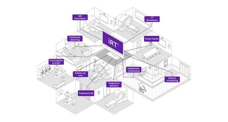
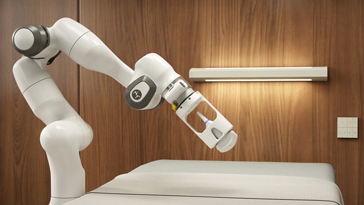
- GE HealthCare Highlights AI-Supported Radiation Therapy Tools at ESTRO 2026
- Nuclear Medicine Set for Continued Growth Driven by Demand for Precision Diagnostics
- GE HealthCare and NVIDIA Collaboration to Reimagine Diagnostic Imaging
- Patient-Specific 3D-Printed Phantoms Transform CT Imaging
- Siemens and Sectra Collaborate on Enhancing Radiology Workflows
 Expo
Expo
- AI Tool Predicts Five-Year Breast Cancer Risk from Mammograms
- AI Mammography Tools Detect Early Breast Cancer Signs Years Before Diagnosis
- Rapid X-Ray Test Quantifies Pulmonary Regurgitation After Tetralogy of Fallot Repair
- AI Tool Flags Osteoporosis Risk from Routine Chest X-Rays
- Simple Chest X-Ray Measure Predicts Survival After Lung Cancer Surgery
- Cardiac MRI Detects Hidden Heart Dysfunction After Heart Attack
- AI Tool Reveals Cortical Lesions on Standard MRI in Multiple Sclerosis
- AI Reconstruction Tool Speeds Dynamic Breast MRI and Improves Cancer Detection
- International Study Assesses AI for Prostate Cancer MRI Interpretation
- AI Approach Could Shorten Advanced Brain MRI Scans by Up to 90%
- Second PSMA PET Identifies Disease in More Than Half of Negative Cases
- New PET Tracer Detects DVT and Pulmonary Embolism in One Scan
- Targeted PET Platform Guides Osteosarcoma Resection and Margin Verification
- Portable PET System Enables Real-Time Bedside Guidance for Biopsies and Ablations
- AI Model Predicts Radiation Dose Before Prostate Cancer Therapy
- Portable 3D Ultrasound System Enables Reproducible Breast Cancer Monitoring
- AI Ultrasound Platform Receives CE Mark for Prenatal Screening
- Hybrid Imaging Platform Reveals How Sleep Supports Brain Waste Removal
- AI Robotic Ultrasound System Automates Echocardiography and Improves Consistency
- Whole Cross-Section Ultrasound System Enables Operator-Independent Imaging
- Virtual Staining Technique Creates Histology Images from CT Data
- CT-Derived Biomarker Predicts Outcomes in Gastric Cancer
- AI Tool Enhances Response Assessment and Survival Prediction in Pleural Mesothelioma
- AI Tool Enables Real-Time Diffuse Optical Tomography for Brain Lesion Detection
- New SPECT/CT Method Differentiates Inflammation from Fibrosis in Interstitial Lung Disease
- Interactive AI Tool Supports Explainable Lung Nodule Assessment
- Breast Imaging Software Enhances Visualization and Tissue Characterization in Challenging Cases
- New Google Cloud Medical Imaging Suite Makes Imaging Healthcare Data More Accessible
- Global AI in Medical Diagnostics Market to Be Driven by Demand for Image Recognition in Radiology
- AI-Based Mammography Triage Software Helps Dramatically Improve Interpretation Process
- GE HealthCare Highlights AI-Supported Radiation Therapy Tools at ESTRO 2026
- Nuclear Medicine Set for Continued Growth Driven by Demand for Precision Diagnostics
- GE HealthCare and NVIDIA Collaboration to Reimagine Diagnostic Imaging
- Patient-Specific 3D-Printed Phantoms Transform CT Imaging
- Siemens and Sectra Collaborate on Enhancing Radiology Workflows

















![Image: (A-C) Representative whole-body maximum-intensity projection images and regional fused PET/CT images from three histologically confirmed osteosarcoma patients who underwent paired [68Ga]Ga-B7-H3-BCH PET/CT and 18F-FDGE PET/CT within 7 days. (D) Multimodal imaging evaluation of patient 3, including X-ray, MRI (T2-weighted imaging, T2WI), CT, and B7-H3 PET/CT. (Photo courtesy of SNMMI)](https://globetechcdn.com/medicalimaging/images/stories/articles/article_images/2026-06-07/294811110_image_1.jpg "Image: (A-C) Representative whole-body maximum-intensity projection images and regional fused PET/CT images from three histologically confirmed osteosarcoma patients who underwent paired [68Ga]Ga-B7-H3-BCH PET/CT and 18F-FDGE PET/CT within 7 days. (D) Multimodal imaging evaluation of patient 3, including X-ray, MRI (T2-weighted imaging, T2WI), CT, and B7-H3 PET/CT. (Photo courtesy of SNMMI)")






 Client Login