Expo
view channel
view channel
view channel
view channel
view channel
view channel
view channel
RadiographyMRI
Nuclear MedicineGeneral/Advanced ImagingImaging ITIndustry News
Events

- AI Technology Predicts Personalized Five-Year Risk of Developing Breast Cancer
- RSNA AI Challenge Models Can Independently Interpret Mammograms
- New Technique Combines X-Ray Imaging and Radar for Safer Cancer Diagnosis
- New AI Tool Helps Doctors Read Chest X‑Rays Better
- Wearable X-Ray Imaging Detecting Fabric to Provide On-The-Go Diagnostic Scanning
- AI Model Outperforms Doctors at Identifying Patients Most At-Risk of Cardiac Arrest
- New MRI Technique Reveals Hidden Heart Issues
- Shorter MRI Exam Effectively Detects Cancer in Dense Breasts
- MRI to Replace Painful Spinal Tap for Faster MS Diagnosis
- MRI Scans Can Identify Cardiovascular Disease Ten Years in Advance
- Novel Bacteria-Specific PET Imaging Approach Detects Hard-To-Diagnose Lung Infections
- New Imaging Approach Could Reduce Need for Biopsies to Monitor Prostate Cancer
- Novel Radiolabeled Antibody Improves Diagnosis and Treatment of Solid Tumors
- Novel PET Imaging Approach Offers Never-Before-Seen View of Neuroinflammation
- Novel Radiotracer Identifies Biomarker for Triple-Negative Breast Cancer
- Breakthrough Deep Learning Model Enhances Handheld 3D Medical Imaging
- Pain-Free Breast Imaging System Performs One Minute Cancer Scan
- Wireless Chronic Pain Management Device to Reduce Need for Painkillers and Surgery
- New Medical Ultrasound Imaging Technique Enables ICU Bedside Monitoring
- New Incision-Free Technique Halts Growth of Debilitating Brain Lesions
- Extending CT Imaging Detects Hidden Blood Clots in Stroke Patients
- Groundbreaking AI Model Accurately Segments Liver Tumors from CT Scans
- New CT-Based Indicator Helps Predict Life-Threatening Postpartum Bleeding Cases
- CT Colonography Beats Stool DNA Testing for Colon Cancer Screening
- First-Of-Its-Kind Wearable Device Offers Revolutionary Alternative to CT Scans
- Global AI in Medical Diagnostics Market to Be Driven by Demand for Image Recognition in Radiology
- AI-Based Mammography Triage Software Helps Dramatically Improve Interpretation Process
- Artificial Intelligence (AI) Program Accurately Predicts Lung Cancer Risk from CT Images
- Image Management Platform Streamlines Treatment Plans
- AI Technology for Detecting Breast Cancer Receives CE Mark Approval
- Patient-Specific 3D-Printed Phantoms Transform CT Imaging
- Siemens and Sectra Collaborate on Enhancing Radiology Workflows
- Bracco Diagnostics and ColoWatch Partner to Expand Availability CRC Screening Tests Using Virtual Colonoscopy
- Mindray Partners with TeleRay to Streamline Ultrasound Delivery
- Philips and Medtronic Partner on Stroke Care

 Expo
Expo
- AI Technology Predicts Personalized Five-Year Risk of Developing Breast Cancer
- RSNA AI Challenge Models Can Independently Interpret Mammograms
- New Technique Combines X-Ray Imaging and Radar for Safer Cancer Diagnosis
- New AI Tool Helps Doctors Read Chest X‑Rays Better
- Wearable X-Ray Imaging Detecting Fabric to Provide On-The-Go Diagnostic Scanning
- AI Model Outperforms Doctors at Identifying Patients Most At-Risk of Cardiac Arrest
- New MRI Technique Reveals Hidden Heart Issues
- Shorter MRI Exam Effectively Detects Cancer in Dense Breasts
- MRI to Replace Painful Spinal Tap for Faster MS Diagnosis
- MRI Scans Can Identify Cardiovascular Disease Ten Years in Advance
- Novel Bacteria-Specific PET Imaging Approach Detects Hard-To-Diagnose Lung Infections
- New Imaging Approach Could Reduce Need for Biopsies to Monitor Prostate Cancer
- Novel Radiolabeled Antibody Improves Diagnosis and Treatment of Solid Tumors
- Novel PET Imaging Approach Offers Never-Before-Seen View of Neuroinflammation
- Novel Radiotracer Identifies Biomarker for Triple-Negative Breast Cancer
- Breakthrough Deep Learning Model Enhances Handheld 3D Medical Imaging
- Pain-Free Breast Imaging System Performs One Minute Cancer Scan
- Wireless Chronic Pain Management Device to Reduce Need for Painkillers and Surgery
- New Medical Ultrasound Imaging Technique Enables ICU Bedside Monitoring
- New Incision-Free Technique Halts Growth of Debilitating Brain Lesions
- Extending CT Imaging Detects Hidden Blood Clots in Stroke Patients
- Groundbreaking AI Model Accurately Segments Liver Tumors from CT Scans
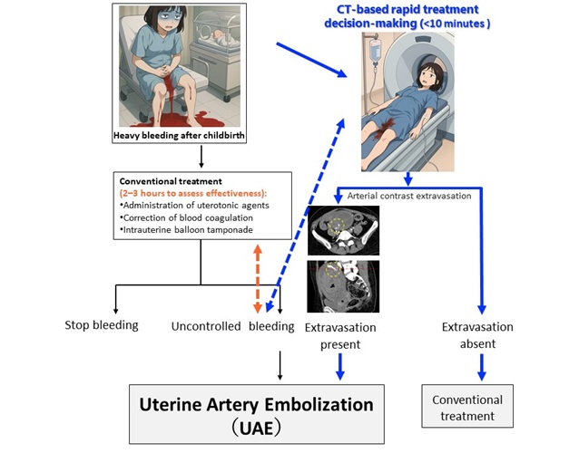
- New CT-Based Indicator Helps Predict Life-Threatening Postpartum Bleeding Cases
- CT Colonography Beats Stool DNA Testing for Colon Cancer Screening
- First-Of-Its-Kind Wearable Device Offers Revolutionary Alternative to CT Scans
- Global AI in Medical Diagnostics Market to Be Driven by Demand for Image Recognition in Radiology
- AI-Based Mammography Triage Software Helps Dramatically Improve Interpretation Process
- Artificial Intelligence (AI) Program Accurately Predicts Lung Cancer Risk from CT Images
- Image Management Platform Streamlines Treatment Plans
- AI Technology for Detecting Breast Cancer Receives CE Mark Approval
- Patient-Specific 3D-Printed Phantoms Transform CT Imaging
- Siemens and Sectra Collaborate on Enhancing Radiology Workflows
- Bracco Diagnostics and ColoWatch Partner to Expand Availability CRC Screening Tests Using Virtual Colonoscopy
- Mindray Partners with TeleRay to Streamline Ultrasound Delivery
- Philips and Medtronic Partner on Stroke Care


 Guided Devices.jpg)


